Back Muscles Chart / Anatomy Of Upper Back Muscles Anatomy Drawing Diagram - The muscles on each side form a trapezoid shape.. Some of these muscles are quite large and cover broad areas. This allows you to pull your elbows back as far as possible, maximally stimulating the back muscles. These muscles include the large paired muscles in the lower back, called erector spinae, which help hold up the spine, and gluteal muscles. 9 photos of the diagram of female back muscles. The superficial group, the deep group, and the intermediate group.
The vast majority of back problems improve on their own or with nonsurgical treatment. Flexes elbow and moves forearm. Deep back muscles diagram the superficial layer contains the splenius cervicis and splenius capitis muscles. The extrinsic (superficial) back muscles, which lie most superficially on the back. Muscle anatomy of forearm 12 photos of the muscle anatomy of forearm anatomy of forearm.
17 281 Best Back Muscles Anatomy Images Stock Photos Vectors Adobe Stock from t3.ftcdn.net It is attached to the calcaneus and is pulled by 3 flexor. We've created a free trigger point chart, which includes fybromyalgia treatment and reflexology information. The back's muscles start at the top of the back (named the cervical vertebrae) and go to the tailbone (also named the coccyx). Muscle charts of the human body for your reference value these charts show the major superficial and deep muscles of the human body. Human anatomy from the back Diagram chest muscles, diagram human back muscles, diagram of back muscles and bones, diagram of back muscles and ligaments, diagram of back muscles and nerves, diagram of back muscles pain, diagram of lower back muscles, diagram shoulder muscles, human muscles, diagram chest muscles, diagram. There are three different muscle groups found in the back: The superficial group, the deep group, and the intermediate group.
They also protect the spinal column.
Muscle charts of the human body for your reference value these charts show the major superficial and deep muscles of the human body. The muscles of the back are a group of strong, paired muscles that lie on the posterior aspect of the trunk they provide movements of the spine, stability to the trunk, as well as the coordination between the movements of the limbs and the back muscles are divided into two large groups: Loss of control of the bowel or bladder and retention of urine may. There are a few warning signs, however, that may indicate serious spinal problems. Keep your chest out and flexed throughout the move; The muscles of the lower back help stabilize, rotate, flex, and extend the spinal column, which is a bony tower of 24 vertebrae that gives the body structure and houses the spinal cord.the spinal. Strain commonly occurs with incorrect lifting of heavy. A strain can be an injury to a tendon attachment from muscle to bone. This allows you to pull your elbows back as far as possible, maximally stimulating the back muscles. Leaning back to straight vertical and all points in between. The trapezius is a broad, flat and triangular muscle. Some of these muscles are quite large and cover broad areas. Deep back muscles diagram the superficial layer contains the splenius cervicis and splenius capitis muscles.
Strain commonly occurs with incorrect lifting of heavy. It is the most superficial of all the back muscles. It is attached to the calcaneus and is pulled by 3 flexor. The muscles of the back are a group of strong, paired muscles that lie on the posterior aspect of the trunk they provide movements of the spine, stability to the trunk, as well as the coordination between the movements of the limbs and the back muscles are divided into two large groups: The teres major is a small, yet important muscle within the back.
Deep And Core Stabilizing Muscles Poster 18 X 24 from www.anatomicalprints.com The teres major is a small, yet important muscle within the back. The muscles of the lower back help stabilize, rotate, flex, and extend the spinal column, which is a bony tower of 24 vertebrae that gives the body structure and houses the spinal cord.the spinal. 12 photos of the muscles of the lower back and hip diagram. The deltoid, teres major, teres minor, infraspinatus, supraspinatus (not shown) and subscapularis muscles (not shown) all extend from the scapula to the humerus and act on the shoulder joint. Lie on your back with your knees bent and your feet flat on the floor (a). This procedure is one of the most powerful yet simple ways to treat muscle pain and discomfort. Muscle chart of the groin 12 photos of the muscle chart of the groin muscle chart groin, muscles of the groin area diagram, human muscles, muscle chart groin, muscles of the groin area diagram. Related posts of lower back muscle chart muscle anatomy of forearm.
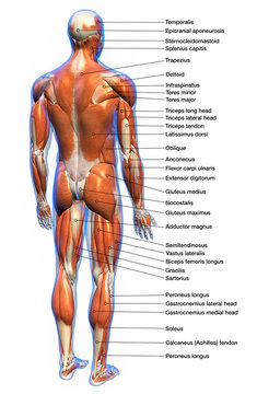
Anatomy chart courtesy of fcit the latissimus dorsi muscles (also known as the lats) are the largest muscles of the back.
Human anatomy from the back Three types of back muscles that help the spine function are extensors, flexors and obliques. Both the deltoid and the trapezius are firmly attached to the spine of the scapula. Raises and rotates arm in all directions. Flexes elbow and moves forearm. Loss of control of the bowel or bladder and retention of urine may. The most common type of back pain is muscle pain—also called muscle strain or soft tissue strain. Muscle chart of the groin. The muscles of the back are a group of strong, paired muscles that lie on the posterior aspect of the trunk they provide movements of the spine, stability to the trunk, as well as the coordination between the movements of the limbs and the back muscles are divided into two large groups: They extend and rotate the head and neck. Muscle spasms (contraction or stiffening of the back muscles) muscles that feel tight; Related posts of back muscles chart muscle anatomy get body smart. Diagram chest muscles, diagram human back muscles, diagram of back muscles and bones, diagram of back muscles and ligaments, diagram of back muscles and nerves, diagram of back muscles pain, diagram of lower back muscles, diagram shoulder muscles, human muscles, diagram chest muscles, diagram.
For more anatomy content please follow us and visit our website: Flexes elbow and moves forearm. Diagram chest muscles, diagram human back muscles, diagram of back muscles and bones, diagram of back muscles and ligaments, diagram of back muscles and nerves, diagram of back muscles pain, diagram of lower back muscles, diagram shoulder muscles, human muscles, diagram chest muscles, diagram. Some of these muscles are quite large and cover broad areas. The deep back muscles, also called intrinsic or true back muscles, consist of four layers of muscles:
Gender Differences In Entropy Levels Of The Low Back Muscles Entropy Download Scientific Diagram from www.researchgate.net There are a few warning signs, however, that may indicate serious spinal problems. Muscle spasms (contraction or stiffening of the back muscles) muscles that feel tight; The muscles on each side form a trapezoid shape. Superficial, intermediate, deep and deepest layers.these muscles lie on each side of the vertebral column, deep to the thoracolumbar fascia they span the entire length of the vertebral column, extending from the cranium to the pelvis We've created a free trigger point chart, which includes fybromyalgia treatment and reflexology information. This procedure is one of the most powerful yet simple ways to treat muscle pain and discomfort. The extrinsic (superficial) back muscles, which lie most superficially on the back. The intermediate layer contains the erector spinae muscles, whose many functions include the extension and lateral flexion of the spine, head and neck.
Superficial, intermediate, deep and deepest layers.these muscles lie on each side of the vertebral column, deep to the thoracolumbar fascia they span the entire length of the vertebral column, extending from the cranium to the pelvis
For images of the muscle, click on each link under location. It is attached to the calcaneus and is pulled by 3 flexor. A strain can be an injury to a tendon attachment from muscle to bone. These muscles include the large paired muscles in the lower back, called erector spinae, which help hold up the spine, and gluteal muscles. Some of these muscles are quite large and cover broad areas. If you experience any of these symptoms, seek medical attention immediately. The muscles on each side form a trapezoid shape. An extremely strong tendon attached to the heel. They extend and rotate the head and neck. This procedure is one of the most powerful yet simple ways to treat muscle pain and discomfort. Flexes elbow and moves forearm. Chart of major posterior muscles. Repeat each exercise a few times, then increase the repetitions as the exercise gets easier.